Etiology of VSD
The definitve cause of any individual congenital heart defect is rarely determined. Congenital heart defects are believed to be multifactorial with both environmental and genetic components.
Genetic risk factors include the presence of certain chromosomal syndromes including: trisomies 13, 18, 21 or a family history of cardiac defects. Environmental factors include: maternal diabetes or phenylketonuria, exposure to disease or teratogens within in the first 8 weeks of gestation. It should be mentioned that a recent study in Northern Ireland found that the risk of VSD was not increased by maternal smoking, alcohol consumption or maternal age. Parent/family education should include the complexity of cardiac development and medical sciences's limited inability to ascertain causality.
Developmental errors
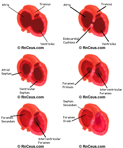
A VSD may result if cells from ventricle, endocardial cushions or truncus arteriosus fail to grow, organize or fuse to the appropriate structures. For example an intact membranous septum requires growth and fusion of tissues derived from the endocardial cushions, the conotruncal ridges and muscular septum. Errors in the growth rate, orientation or fusion of any of these tissues can result in VSD.
 Endocardial cushions
The first division is accomplished by the endocardial cushions. The endocardial cushions begin the separation of the heart into right and left, upper and lower chambers. These chambers will become the atria and ventricles. While the endocardial cushions continue to develop, the atrial and ventricular septa begin to form.
Muscular Septum
The ventricular septum is composed of thick, trabeculated muscular tissue derived from the walls of the growing ventricles. When this muscular septum is complete, a large opening (the interventricular foramen) still remains between the two ventricles. The interventricular foramen is usually completely closed by week seven. Closure is accomplished by growth of membraneous tissue derived from the endocardial cushions, the interventricular septum and from the conus ridges growing within the truncus.
Membranous septum
The membranous septum derived from tissue growing within the truncus. The truncus begins to divide about 29 days after conception. The division starts with the growth of two ridges that take a spiral path toward the ventricles. The spirals eventually joining with tissue from the interventricular septum and closing the interventricular foramen. This tissue completes the separation of venous from arterial blood flow.
RnCeus
Homepage | Course
catalog | Discount
prices | Login | Nursing
jobs | Help
2007 ©RnCeus.com