Anatomic Landmarks
It is important to review the anatomy of the chest wall and thoracic cavity, as you will use anatomic landmarks to document the location of respiratory assessment findings.
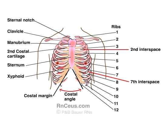
The thoracic cavity is made up of 12 pairs of ribs that connect in the posterior thorax to the vertebral bodies of the spinal column. In the anterior thorax, the first 7 pairs of ribs are attached to the sternum or breastbone by cartilage. The lower 5 ribs do not attach to the sternum. The 8th, 9th, and 10th ribs are attached to each other by costal cartilage. The 11th and 12th ribs, known as “floating ribs,” are not attached in any way to the sternum; they move up and down in the anterior chest, allowing for full chest expansion.
Please review the important landmarks of the bony thoracic anatomy.
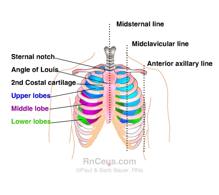
The following diagram shows the anterior chest again, with the lobes of the lungs included. Various reference lines and angles are commonly used to identify respiratory findings. For example:
Other terms used to document locations for chest physical assessment include:
© RnCeus.com