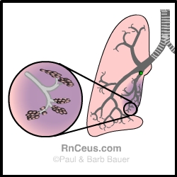
Atelectasis: In this condition, an area of the lung or an entire lung collapses. Atelectasis may be due to airway obstruction, or compression of the lung. In the diagram above, an obstruction blocks the airway, causing the associated alveoli to collapse and that area of the lung to shrink. Any alveolar air beyond the obstruction becomes absorbed by the pulmonary capillaries, and the alveolar walls cave in.
| Assessment findings include: | |
|
Inspection |
|
|
Palpation |
|
|
Percussion |
|
|
Auscultation |
|
To see a slide of severe atelectasis click on this URL http://www-medlib.med.utah.edu/WebPath/LUNGHTML/LUNG188.html