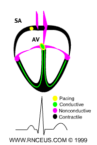
|
Normal
cardiac conduction occurs as:
- an impulse is generated at the SA node and
spreads across both atria, causing them to
contract. Note that the atrial impulse correlates
with the P wave in the EKG diagram.
- The Fibro-fatty atrioventricular groove
insulates the ventricles from the atrial impulse.
The AV node is the only normal gateway of
conduction to the ventricles.
- The impulse is delayed at the AV node, travels
down the AV bundle and it's branches and
reaches the Purkinje fibers. The
ventricles are stimulated to contract. Follow the
red dots and note correlation with the QRS
wave in the EKG diagram above.
- The T wave correlates with repolarization
of the ventricles.
|