Blood
Flow: Review
In order to grasp the concepts
of measuring and interpreting hemodynamic values, it is important to understand
how blood flowing through the heart is related to the cardiac cycle.
|
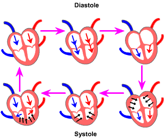
Diastole:
The myocardium is relaxed. The atria and ventricles fill
passively. AV valves allow blood to pass from the atria to the ventricles. The aortic and
pulmonary artery semilunar valves are closed because the blood in those
vessels is at a higher pressure than the ventricles. Blood continues
to fill atria and ventricles, stretching the compliant heart cells.
Systole:
- The atria contract
and eject the final amount of blood into the ventricles. The atrial contraction contributes only about 10% to the total ventricular
volume, while the patient is at rest. If the heart rate is high and the ventricles don't have
time to fill completely, atrial systole can contibute as much as 40%.
- Atria relaxation
causes atrial pressure to be lower than ventricular pressure.
- High ventricular
pressure relative to the atria causes the AV valves to close, preventing
backflow while the ventricles contract.
- The ventricles
continue to contract, ejecting blood through the semilunar valves out
to the lungs and rest of the body.
|
 |
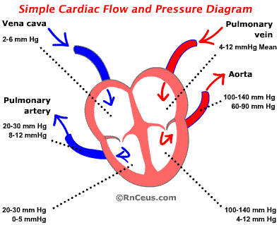 Fluid
flows from high pressure to lower pressure.
Fluid
flows from high pressure to lower pressure.
Blood within the cardiovascular system adheres to this rule. This is evidenced
by the direction of blood flow. The higher pressure generated by the left heart
produces a gradient which moves blood from the left heart, through the body
and into the right side of the heart.
When the left ventricle
(LV) contracts, it generates a systolic blood pressure of 100-140 millimeters
of Hg (mm Hg).
- The aortic diastolic
pressure is usually 60-90 mm Hg. The LV/aortic pressure gradient causes blood
to pass through the aortic valve.
- Blood flowing from the
LV to the aorta raises the aortic pressure to equal the LV pressure.
- A momentary aortic systolic
pressure of 100-140 mm Hg is then dissipated across the capillary beds.
- Capillary pressure exceeds
that of the venuoles. The capillary/venuole gradient causes blood to flow
into the low pressure venous system.
- Low pressure venous blood
is returned to the right atrium, aided by skeletal muscle compression, negative
intra-thoracic pressure and a multitude of one-way valves that advance the
blood toward the vena cavae.
The
pressure of blood within the right atrium is the central venous pressure (CVP). The blood pressure of the vena cavae is similar to the CVP because there are
no valves or flow obstructions between the vena cavae (VC) and the RA. The VC
and heart's right side can be viewed as one chamber with a contractile portion
at the distal end. The CVP averages between 2-6 millimeters of mercury (mm Hg).
During right ventricular
(RV) diastole, the pressure within the RV is between 0-5 mm Hg. Elasticity
and compliance of the ventricular myocardium help generate a lower intraventricular
pressure. Lower intraventricular pressure, aided by atrial systole, causes blood
to flow across the open atrioventricular AV valve.
Right
ventricular systolic pressure is usually from 20-30 mm Hg.
This exceed the right atrial pressure. The pressure gradient applies greater
pressure to the ventricular side of the AV valve, which causes it to close.
The pulmonary artery
(PA) pressure,
prior to systole, is normally 8-12 mm Hg.
During RV systole the PA pressure will rise to equal the RV pressure, usually
20-30 mm Hg. The systolic PA pressure of 20-30 Hg is quickly dissepated by the
compliance of the pulmonary vascular bed to a diastolic pressure of 8-12 Hg.
Blood leaves the pulmonary
vasculature at about 4-12 mm Hg, passively entering the pulmonary veins.
The pulmonary veins empty directly into the left atrium. Elasticity and compliance
of the ventricular myocardium help generate a slightly lower intraventricular
filling pressure. Lower intraventricular pressure, aided by atrial systole,
causes blood to flow across the open atrioventricular AV valve.
LV systole generates 100-140 mm Hg. Aortic diastolic pressure is usually
60-90mm Hg. The pressure gradient of 100-140/60-90 mm Hg drives blood into the
aorta and onward to the rest of the body. The cycle is complete.
Instant
Feedback:
What
causes the AV valve to close.