Physiology of Cardiac Conduction
 A healthy heart is capable of altering cardiac output to match the physical demands of the body. The normal adult resting heart rate is between 60-100 beats per minute (bpm) but when stressed or exercising the same heart may rapidly increase to a rate of 140 bpm or more. The change in rate (chronotropism), contractility (inotropism), and conduction (dromotropism) are regulated by the autonomic nervous system.
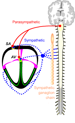
The supraventricular heart is innervated by both sympathetic and parasympathetic (vagal) fibers of the autonomic nervous system. The ventricular actions are primarily modulated by sympathetic influence. Sympathetic stimulation of cardiac β1/β2 receptors by the neurotransmitter norepinehrine and circulating adrenal epinephrine increase cardiac chronotropy, inotropy and dromotropy. Parasympathetic (vagal) stimulation of muscarinic M2-receptors by the neurotransmitter acetlycholine (ACh) has an opposite effect. The autonomic nervous system system modifies cardiac output by extrinsic stimulation.
Although the autonomic system regulates extrinsic aspects of cardiac output, each cardiac myocyte has the intrinsic ability auto-depolarize, i.e. to self-initiate a change of electrical charge. This characteristic is known as automaticity and it is confined to cells in the heart and brain. In the heart, the sinoatrial node (SA) is a collection of specialized myocytes that are able to auto-depolarize and repolarize faster than any other myocytes.
The Sinoatrial node (SA) sets the heart rate. It is the "pacemaker" because its fastest ability to auto-depolarize and repolarize makes it the first to initiating each normal heart beat. It is believed that initial SA node action potential propagates from a small group of cells near the center of the SA node. The SA node is supraventricular and is sensitive to parasympathetic and sympathetic influence.
The atrioventricular node (AV) and the Bundle of His/Purkinje fibers are two additional groups of specialized pacemaker cardiomyocytes, they have the second and third fastest automaticity rates respectively. If the SA node fails to initiate AV depolarization, the AV node takes over the role of ventricular pacer. The Bundle of His/Purkinje fibers are the slowest self-depolarizing pacemaker cells, they become the ventricular pacemaker of last resort. Finally, healthy contractile myocytes have the necessary equipment for automaticity but they require a much greater negative resting voltage to initiate a spontaneous action potential.
Pacemakers myocardial cells depolarize and repolarize in a series of electrical phases. Each phase reflects the staged movement of ions (principally Na+, Ca++ and K+) into (influx) or out (efflux) of the pacemaker cell. The ions move across the cell membrane through ion channels. Some channels are always open and their specific ions can move freely from high concentration to low concentration. Other voltage-gated channels open and close depending upon internal/external voltage differential. When a channel is opened, ions specific to that ion channel move from high concentration to low concentration. Closure of an ion channels decrease ion influx or efflux. As ions flow through open channels, they alter the distribution of charge across the membrane. In addition, like all cells pacemaker cells utilize ATP powered pumps to actively concentrate in the cytosol or remove ions from the cell.
When a SA pacer cell depolarizes, the voltage change generates an action potential that depolarizes connected cells in a chain reaction that spreads via gap junctions, throughout the cardiac conduction system, and onward to all the cardiac myocytes, causing atrial and then ventricular contraction. Retrograde conduction does not normally occur because a depolarized cell requires time (refractory period) to redistribute ions (repolarize) before it can be depolarized.
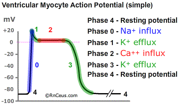
Ventricular cardiomyocytes work to acheive a stable resting hyperpolarized (-) internal charge, known as Phase 4. They do this by pumping Na+and Ca++ ions out of the cell and K+ into the cytosol. Contractile myocytes maintain a relatively negative internal charge until they are depolarized by a sufficient stimulus.
The stimulus is usually caused by the depolarization of a neighboring cardiomyocyte. High internal concentration of Na+ and Ca++ lons within the depolarized neighboring cell cause some ions to leak through the gap junctions connecting cells. The leaked Na+ and Ca++ions raise the internal potential to the threshold potential of about -70mV.
 When the stimulus is sufficient:
- Phase 0 begins at the Threshold -70mV; Na+channels open and Na+ions rush into the cell. This Na+ influx rapidly drives the (-) resting internal charge to (0mV) state of depolarization and a brief overshot (+) charge. Slow Ca++ channels open around -40mV allowing a slow influx of Ca++.
- Phase 1 begins when voltage gated K+channels open allowing K+ ion efflux while continued influx of Ca++ decreases the internal (+) charge back to about (+0-).
- Phase 2 begins when the slow Ca++ influx concentration triggers a much greater release of Ca++ ions stored in the sarcoplasmic reticulum (SR). The SR Ca++ release induces cardiac myofibril contraction. Contraction begins about halfway through Phase 2. Phase 2 plateaus for about 200 milliseconds, after which contraction stops, and the Ca++ channels are closed.
- During Phase 3 (repolarization), K+ channels continue to efflux, making the internal charge more (-).
- Phase 4 hyperpolarization, (-) resting state is acheived by ATP powered outward pumping of Na+ and Ca++ ions as well as recharging the SR Ca++. The (-) charge is maintained by (Na+/K+)ATPase that pumps 3Na+ ions out for every 2K+ ions it brings into the cell.
In contrast to ordinary cardiac myocytes, pacemaker cells do not sustain a stable
Phase 4 (-) internal charge. Instead, as soon as pacemaker cell reaches hyperpolarization the internal voltage slowly begins to become more positive.
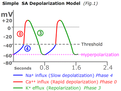 Phase 4, the internal (-) charge slowly begins to become more (+) as soon as the cell reaches an internal charge of -50 mV or greater (hyperpolarization). The slow diastolic depolarization of pacemaker cells is the hallmark of automaticity. Pacemaker automaticity is due to "funny channels" that allow the slow influx of Na+until the threshold voltage is met.
- Phase 0 - At threshold voltage, Ca++ channels open and Ca++ ions flood into the cytosol. Ca++ ions quickly raise the internal charge to (+) depolarizing the pacer cell. Depolarization initiates an electrical wave of depolarization that spreads from one pacer cell to another. Depolarization closes the Ca++ channels.
- Phase 3 - Repolarization begins as K+ channels open allowing the positively charge K+ ions to exit while the Na+ and Ca++ ion pumps work to remove those ions and return the internal charge toward the negative hyperpolarized state.
The flow of ions through membrane channels and the ion exchange/pumping processes influence the threshold, slope, amplitude and duration of depolarization and repolarization contraction cycle. The sinoatrial pacemaker regulates heart rate which is largely influenced by the slope of phase 4. Parasympathetic acetylcholine increases the time to threshold and catecholamines decrease the time to threshold.
Instant Feedback:
Parasympathetic acetylcholine causes muscarinic receptors to
References:
Pollock, J. D., & Makaryus, A. N. (2022). Physiology, Cardiac Cycle. In StatPearls. StatPearls Publishing.
Wei, X., Yohannan, S., & Richards, J. R. (2023). Physiology, Cardiac Repolarization Dispersion and Reserve. In StatPearls. StatPearls Publishing.