
| Rate |
Atrial 250-350/min; ventricular conduction depends on the capability of the AV junction (usually rate of 150-175 bpm). |
| P wave | Not present; usually a saw tooth; pattern is present. |
| QRS | Normal |
| Conduction | 2:1 atrial to ventricular most common. |
| Rhythm | Usually regular, but can be irregular if the AV block varies. |
| Symptoms | Palpitations, rapid heart rate, chest pain, shortness of breath, lightheadedness, fatigue, and low blood pressure. |
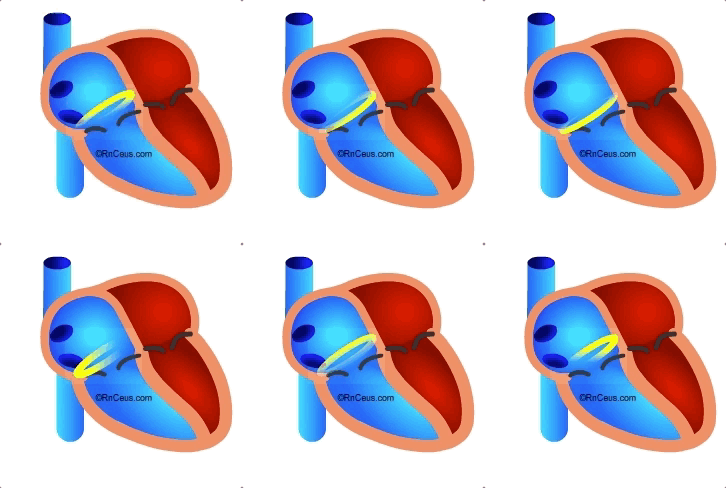
Atrial flutter is the second most common tachyarrhythmia, after atrial fibrillation. It is usually confined to tissue of the right atrium, only rarely passing through the atrial septum to effect the left atrium. It results from an aberrant conduction circuit typically located in the tissue between the inferior vena cava and the tricuspid valve, an area known as the cavotricuspid isthmus. This area is contains many intersecting fibers which may increase the chance of aberrant conduction. The conduction almost always follows in a counter-clockwise path through the isthmus and across the walls of the right atrium.
 Atrial flutter almost always occurs in diseased hearts but it can occur in otherwise asymptomatic hearts. The incidence of atrial flutter increases with age and medical conditions including: congestive heart failure, rheumatic valve disease, congenital heart disease, lung disease such as emphysema, or high blood pressure. Surgery involving the right atrium can increase the risk of atrial flutter due to scar formation in the atrial wall or tricuspid annulus.
Treatment depends on the level of hemodynamic compromise.
Instant Feedback:
© RnCeus.com