Partitioning of Truncus Arteriosus
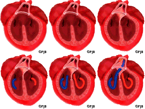
 The truncus begins to divide about about week 5. The division starts with the growth of two ridges derived from the limbs of the cardiac crescent. The ridges continue to grow in a spiral path toward the ventricles, eventually joining with tissue from the interventricular septum and closing the interventricular foramen. This tissue completes the separation of venous from arterial blood flow.
The processes of septation and spiraling divide the truncus lumen into the proximal ascending aorta and the pulmonary trunk. Fusion of the truncal and conal swellings establishes the right ventricular origin of the pulmonary trunk and left ventricular origin of the aorta. The aortopulmonary valves develop at the lines of fusion of truncal and conal swellings. Spiralling of the septum allows the pulmonary trunk to cross anterior and to the left of the aorta (Bhatia, 2022).
References
Bhatia, S., Munir, S., Aly, A. (n.d.). Pediatric cardiology part 1. Embryology.
Retrieved July 9, 2022, from https://www.utmb.edu/pedi_ed/CoreV2/CardiologyPart1/CardiologyPart13.html
 The truncus begins to divide about about week 5. The division starts with the growth of two ridges derived from the limbs of the cardiac crescent. The ridges continue to grow in a spiral path toward the ventricles, eventually joining with tissue from the interventricular septum and closing the interventricular foramen. This tissue completes the separation of venous from arterial blood flow.