Hemoglobin
 Hemoglobin
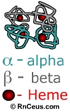
is a molecule comprised of four subunits. Each subunit contains an iron containing
pigment (heme) and a protein (globulin). There are two types of subunits, alpha
and beta. Each gram of hemoglobin can carry 1.34 ml of oxygen. The oxygen carrying
ability of blood is directly proportional to its hemoglobin concentration. The
numbers of red blood cells does not indicate blood's oxygen content because
some cells may contain more hemoglobin than others. Hemoglobin determination
is used to screen for anemia, to identify the severity of anemia, and to assist
in evaluating the patient's response to anemia therapy. Hemoglobin also serves
as an important pH buffer in the extracellular fluid.
Hemoglobin
is a molecule comprised of four subunits. Each subunit contains an iron containing
pigment (heme) and a protein (globulin). There are two types of subunits, alpha
and beta. Each gram of hemoglobin can carry 1.34 ml of oxygen. The oxygen carrying
ability of blood is directly proportional to its hemoglobin concentration. The
numbers of red blood cells does not indicate blood's oxygen content because
some cells may contain more hemoglobin than others. Hemoglobin determination
is used to screen for anemia, to identify the severity of anemia, and to assist
in evaluating the patient's response to anemia therapy. Hemoglobin also serves
as an important pH buffer in the extracellular fluid.
- Normal
hemoglobin values are:
- Adult:
(males): 13.5 - 17 g/dl
- (Females):
12 - 15 g/dl
- Pregnancy:
11 - 12 g/dl
- Newborn:
14-24 g/dl 77% of this value is
fetal hemoglobin, which drops to approximately 23% of the total at 4
months of age
- Children:
11-16 g/dl
|
Glucose
irreversibly attaches to hemoblobin and other proteins that on contacts.
Measurement
of hemoglobin A-1C or glycosylated hemoglobin is used
to monitor and evaluate diabetes. The hemoglobin A-1C
reflects an average blood glucose over a 3 month period, compared to a
fasting blood glucose that reflects blood glucose during a one-time fasting
state. Adult non-diabetics have a hemoglobin A-1C value
between 2% and 5%. Diabetics with effective disease control have hemoglobin
A-1C values between 2.5% and 6%. Diabetics with poor disease control may have values of 8% and higher.
|
Decreased hemoglobin:
Blood loss and bone marrow
suppression reduce total RBC count and therefore lower total hemoglobin content.
Hemoglobin levels are also lowered in patients who have abnormal
types of hemoglobin or hemoglobinopathies. Red blood cells with abnormal types
of hemoglobin are often fragile and damaged or destroyed easily in the vascular
system. Hemoglobin electrophoresis can distinguish among specific types of abnormal
hemoglobin.
Thalassemia is an inherited
recessive hemoglobinopathy. It results from a failure to produce sufficient
globin molecules. The failure can be in either the alpha or beta portion. In
sickle cell anemia, the patient has an abnormally shaped hemoglobin known as
sickle hemoglobin (hgbS). Sickle hemoglobin creates misshapen RBCs which form
blockages in the vessels.
Other patients have a normal
RBC count but a low hemoglobin level. This situation occurs with iron-deficiency
anemia, in which red blood cells have less hemoglobin than normal. Iron deficiency
anemia is also referred to as hypochromic anemia. Hypochromic is a term that
means "less than normal color." In general, women need more iron in
their diets than men, due to the regular loss of iron in the menstrual flow.
During pregnancy a woman's need for iron to build more hemoglobin increases.
If a woman becomes pregnant when she has low iron reserves, she is at risk of
becoming severely anemic. Regular hemoglobin testing is an important part of
prenatal care. During the last trimester of pregnancy, a condition known as
"physiological anemia of pregnancy" occurs. This normal drop in hemoglobin
values results from an increase in the plasma volume. Multiple blood draws in
premature infants is a common cause of anemia.
Instant
Feedback:
Red
blood cells that have abnormal hemoglobin are damaged or destroyed more easily
than cells with normal hemoglobin.
Hemoglobin: critical low
and high values
- A hemoglobin value under
5 g/dl may cause
heart failure
- A hemoglobin value over
20 g/dl may cause clogging of capillaries
due to hemoconcentration
Increased levels of hemoglobin
are found in any condition in which the number of circulating red blood cells
rises above normal. Examples of conditions associated with increases in hemoglobin
are polycythemia vera , severe burns, chronic obstructive pulmonary disease,
and congestive heart failure.
For more
information about anemia, consider visiting the AMERICAN SOCIETY of HEMATOLOGY
http://www.hematology.org/Patients/Other-Resources/Education-Book/5302.aspx
© RnCeus.com